

Ultrasound Sticker for Continuous Imaging
MIT engineers designed an adhesive patch that produces ultrasound images of the body. The stamp-sized device sticks to skin and can provide continuous ultrasound imaging of internal organs for 48 hours. Image: Felice Frankel
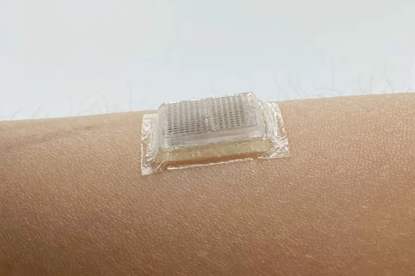
Researchers at MIT have developed a bioadhesive ultrasound device that when adhered to the skin can possibly replace (or augment) ultrasound imaging wands. Image: MIT
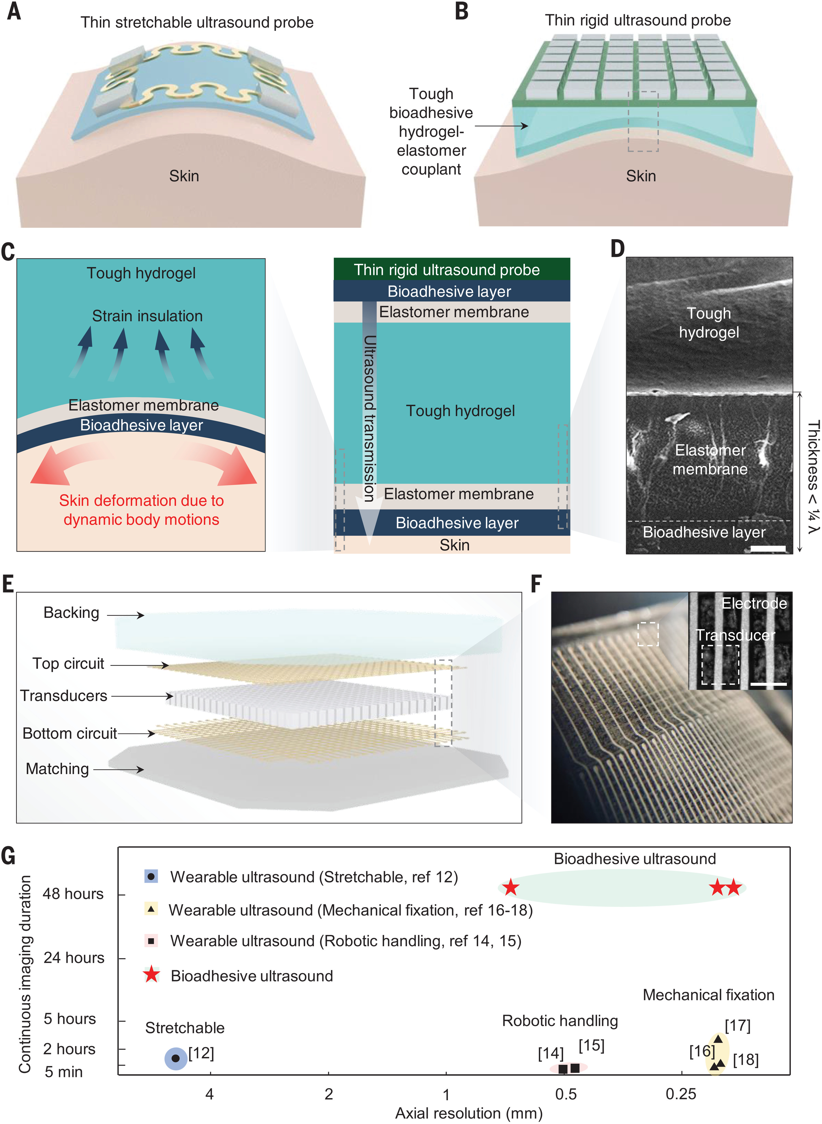
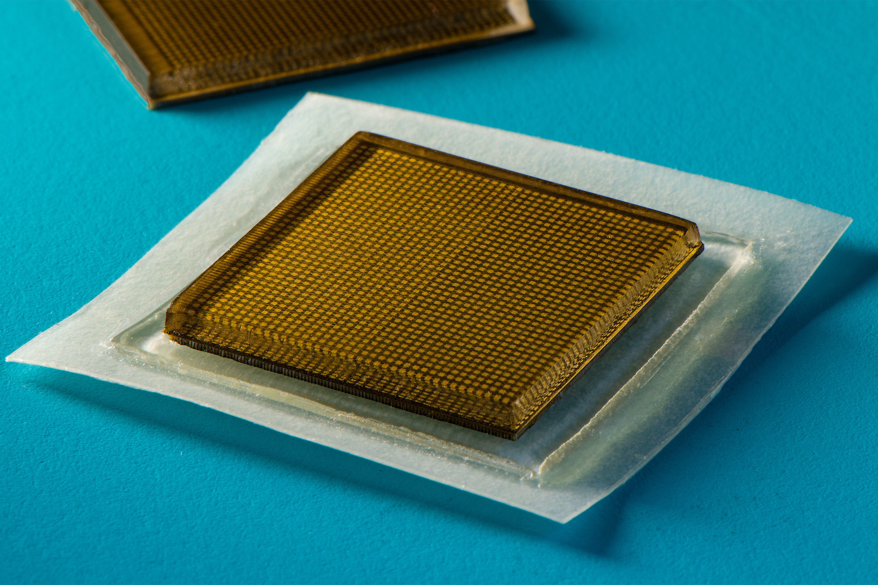
Design and imaging performances of the BAUS device — (A) Existing epidermal wearable ultrasound imaging usually relies on thin and stretchable devices physically attached to the skin. (B) The BAUS device consists of a thin and rigid ultrasound probe robustly adhered to the skin via a couplant made of a soft, tough, antidehydrating, and bioadhesive hydrogel-elastomer hybrid. The dashed rectangle indicates the region shown on the right in (C). (C) The BAUS couplant consists of a tough hydrogel, containing 90 wt % water, that is encapsulated by an elastomer membrane further coated by a thin bioadhesive layer. The BAUS couplant maintains robust adhesion between the BAUS probe and the skin over 48 hours and insulates the BAUS probe from skin deformation during dynamic body motions. (D) Scanning electron microscopy (SEM) image of the cross section of the BAUS couplant. The total thickness of the elastomer membrane and the bioadhesive layer is less than one-fourth of the ultrasound wavelength λ. Scale bar is 10 μm. (E) Schematics of the BAUS probe structure. The BAUS probe consists of an array of high-performance piezoelectric elements, which are controlled by the top and bottom circuits. The top and bottom circuits are covered by the acoustic backing and matching layers, respectively. The dashed rectangle indicates the region shown in (F). (F) Optical microscopic image of a BAUS probe with 3D-printed top and bottom circuits. The inset image provides a zoomed-in top view of the device showing the top circuit and piezoelectric elements. Scale bar is 0.5 mm. (G) Comparison of axial-imaging resolutions and continuous-imaging durations of existing wearable ultrasound imaging devices and the BAUS device. Image: Chonghe Wang et al., Bioadhesive ultrasound for long-term continuous imaging of diverse organs. Science 377, 517-523 (2022).