

Nanoscale modifications yield a 10x enhancement in scintillator efficiency
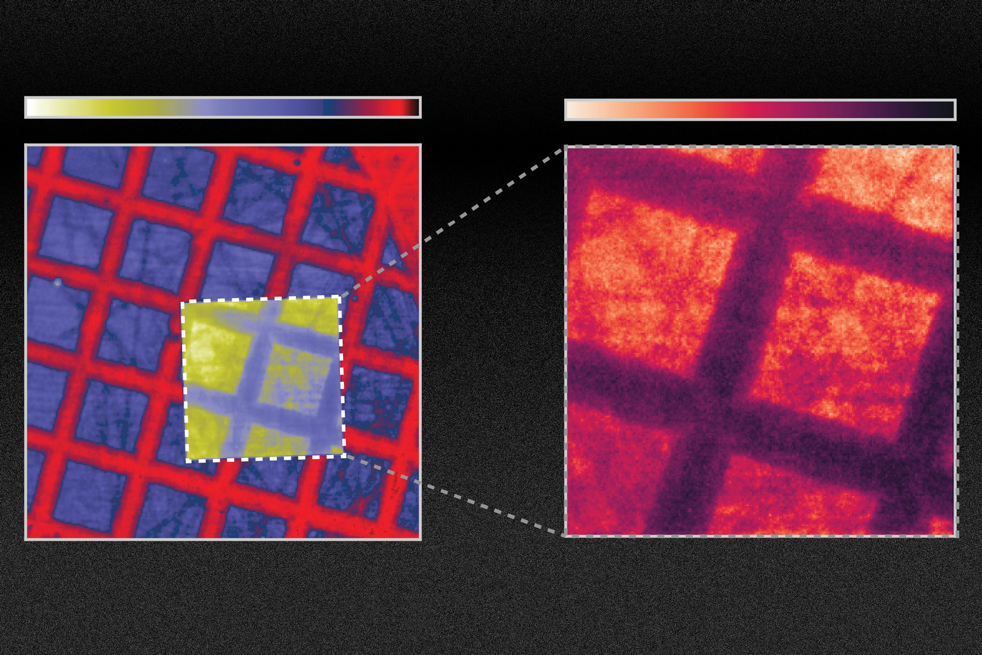
Researchers at MIT have shown how one could improve the efficiency of scintillators by at least tenfold by changing the material’s surface. This image shows a TEM grid on scotch tape, with the right side showing the scene after it is corrected. Image: Courtesy of the researchers, edited by MIT News
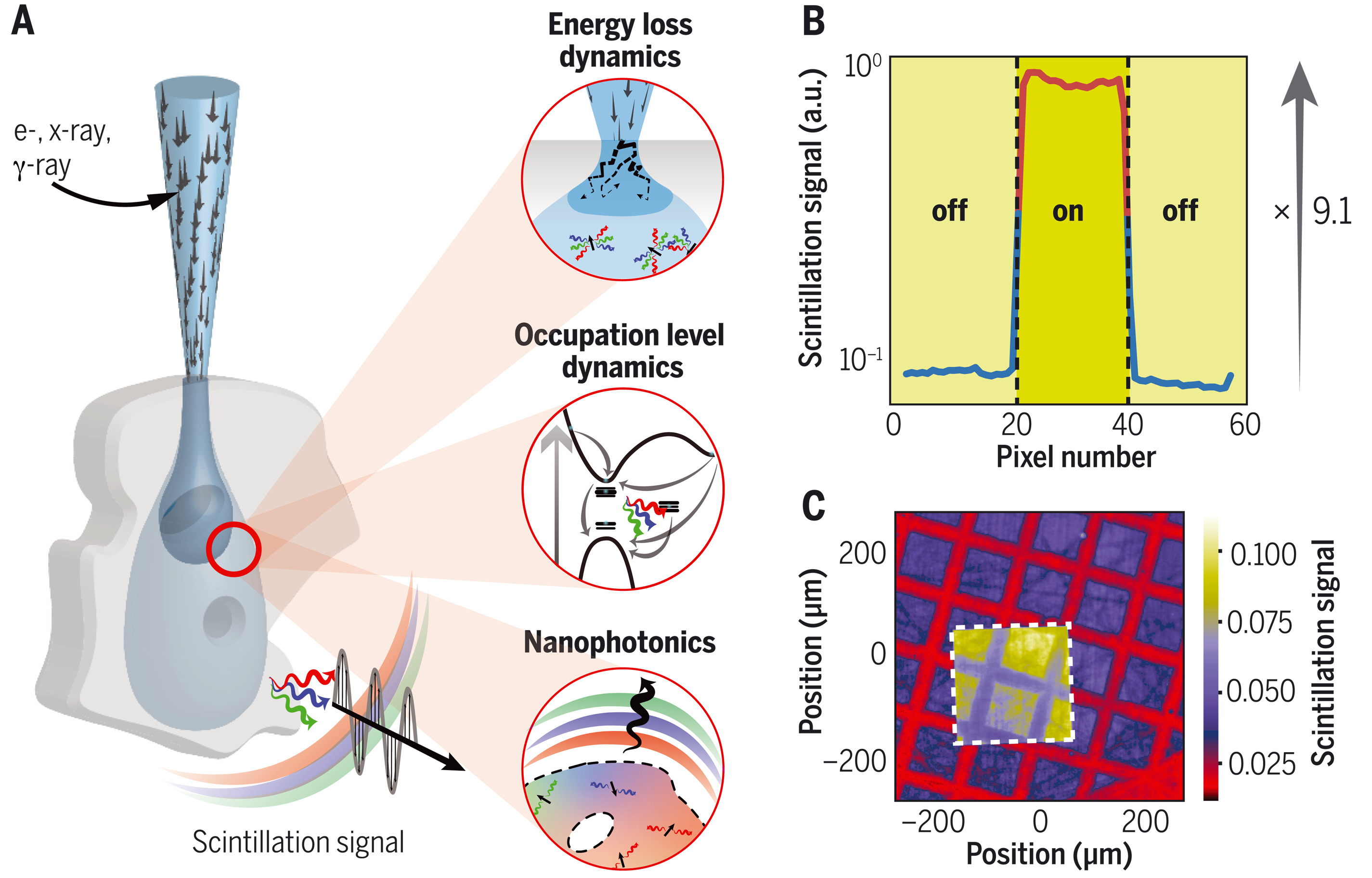
Nanophotonic scintillators — (A) Nanophotonic scintillators consist of nanophotonic structures integrated with scintillators. Scintillation can be modeled, tailored, and optimized by combining energy loss dynamics, occupation level dynamics, and nanophotonics modeling. (B) Order-of-magnitude x-ray scintillation enhancement with a photonic crystal nanophotonic scintillator. (C) X-ray scan taken with a nanophotonic scintillator (white dashed square). Image: Charles Roques-Carmes et al., A framework for scintillation in nanophotonics. Science 375, eabm 9293 (2022)