Nanoparticle-Based Onco-Fluorescent Agents

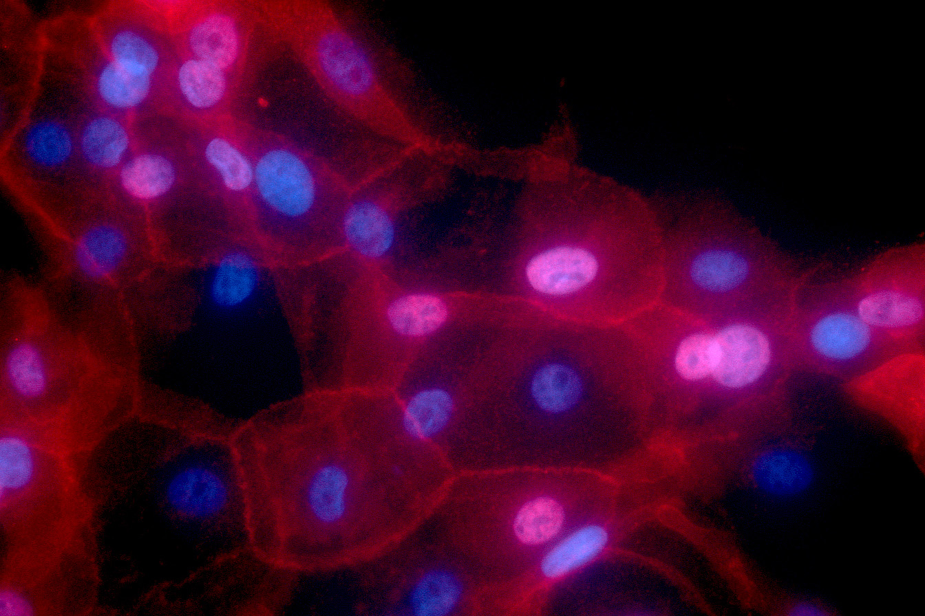
Lumicell has developed a handheld device and an optical imaging agent that allow surgeons to scan the tissue within the surgical cavity to visualize residual cancer cells. Image: National Institutes of Health
Leveraging ISN-derived nanoparticle technology, spinoff company Lumicell has received FDA approval for the LumiSight system, which targets a tumor microenvironment and fluoresces near cancer cells at the tumor margin, for the treatment of breast cancer. This enables a surgeon to view tumor cells that are not otherwise visible to the naked eye, reducing the likelihood of a second surgery.
The imaging agent—a fluorophore dye tethered by peptides to a molecular quencher—is intravenously administered. Enzymes secreted by cancerous cells cleave the peptide links, freeing the dye and causing it to glow in proximity to the cancer when viewed with the Lumicell Direct Visualization System (DVS), a handheld, single-cell-resolution imaging device. Software produces real-time images of the potential location of residual cancer, adding an average of less than seven minutes to the surgical procedure.
In a clinical trial of more than 350 patients, Lumicell’s technology not only reduced the need for second surgeries but also detected cancerous tissue that standard pathology might miss.
In addition to its approval for use with breast cancer, feasibility studies are underway for the system’s use with other cancers, such as gastrointestinal (GI) cancers, while exploratory work is in progress for use in the treatment of sarcoma, brain, and prostate cancers.

